46
Onco
l
Vol 7
l
N°2
l
2013
quences de la résécabilité (17), celle-ci étant prédictive de la
survie, tandis que la nécessité de reconstruction artérielle est
généralement synonyme de mauvais pronostic (18).
Il y a donc intérêt à évaluer correctement le stade d'envahisse-
ment vasculaire avant chirurgie, ce que l'échographie par voie
endoscopique semble permettre de manière plus efficace que
le CT-scan (qui reste le standard) en cas de doute (19). Cepen-
dant, avant de conclure fermement, il faut se rappeler que les
études comparatives en la matière sont très hétérogènes (20).
Détecter les métastases
L'envahissement ganglionnaire doit également être établi avec atten-
tion, car le pronostic des patients N3 est équivalent à celui des pa-
tients en phase métastatique. Ils ne doivent donc pas être opérés.
Côté hépatique, la technique actuelle de choix est l'imagerie de
résonance en diffusion pondérée, meilleure que le scanner
multibarrettes (21). Quant à l'échoendoscopie, elle permet de
détecter certaines lésions occultes dans le lobe gauche hépa-
tique (31%) et en cas de carcinomatose avec ascite (40%) (22).
Enfin, une méta-analyse datant de 2011 a montré que si
l'échoendoscopie est très spécifique, le PET-scan au 18F-FDG
est très sensible (23), du moins pour le stade III (24). Il n'est
cependant pas stricto sensu une aide au diagnostic, notamment
différentiel entre masse inflammatoire et néoplasique.
Au niveau biologique, le taux de CA19-9 est plus pronostique
que diagnostique (25).
The tissue is the issue: refrain connu?
Dans un futur proche, la prise en charge d'une lesion néopla-
sique nécessitera l'obtention de tissu à visée non seulement
diagnostique, mais également pronostique et prédictive.
Au départ d'une même quantité de tissu, plusieurs méthodes,
telles que la méthode FISH et l'activité de la télomérase, per-
mettent de détecter les anomalies chromosomiques (27) et d'affi-
ner le diagnostic de malignité. Quant à la prédictivité de la réponse
à la chimiothérapie, elle peut être affinée par l'évaluation de
hENT1, dont l'expressivité élevée est associée à un taux de survie
meilleur en cas de traitement par gemcitabine post-résection (28).
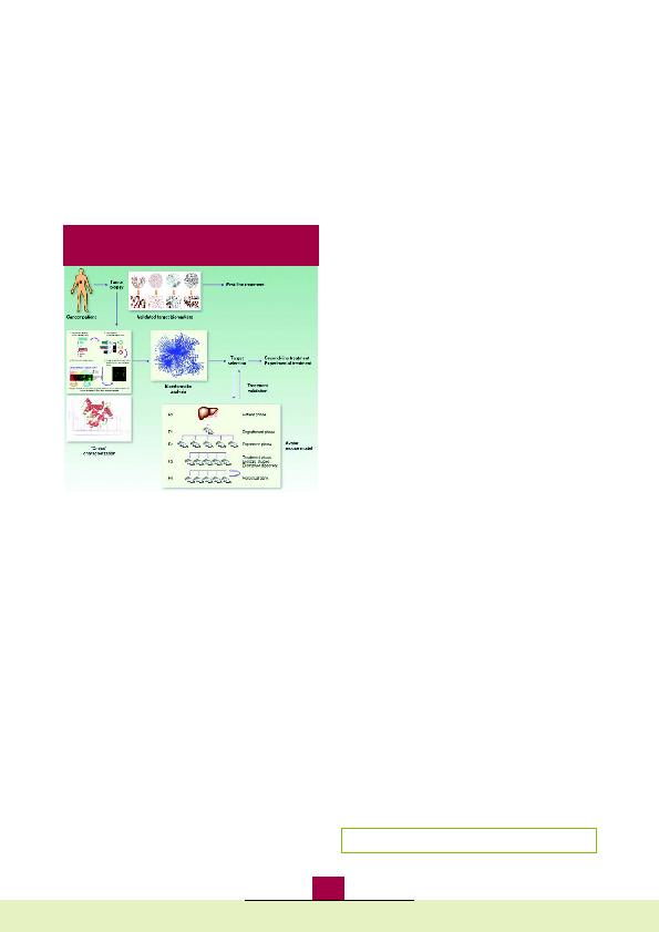
L'aspiration à l'aiguille fine écho-endoscopiquement guidée
pourrait aussi affiner le pronostic et guider le traitement néo-
adjuvant ou le choix du traitement dans le futur (Figure 2).
Références
1.
Low G, Panu A, Millo N, Leen E. Multimodality imaging of neoplastic and nonneoplastic solid
lesions of the pancreas. Radiographics 2011;31(4):993-1015.
2.
Noone TC, Hosey J, Firat Z, Semelka RC. Imaging and localization of islet-cell tumours of the
pancreas on CT and MRI. Best Pract Res Clin Endocrinol Metab 2005;19(2):195-211.
3.
Ichikawa T, Sou H, Araki T, et al. Duct-penetrating sign at MRCP: usefulness for differentiating
inflammatory pancreatic mass from pancreatic carcinomas. Radiology 2001;221(1):107-16.
4.
Siddiqi AJ, Miller F. Chronic pancreatitis: ultrasound, computed tomography, and magnetic
resonance imaging features. Semin Ultrasound CT MR 2007;28(5):384-94.
5.
Tajima Y, Kuroki T, Tsutsumi R, Isomoto I, Uetani M, Kanematsu T. Pancreatic carcinoma
coexisting with chronic pancreatitis versus tumor-forming pancreatitis: diagnostic utility of
the time-signal intensity curve from dynamic contrast-enhanced MR imaging. World J
Gastroenterol 2007;13(6):858-65.
6.
Bali MA, Metens T, Denolin V, et al. Tumoral and nontumoral pancreas: correlation between
quantitative dynamic contrast-enhanced MR imaging and histopathologic parameters.
Radiology 2011;261(2):456-66.
7.
Klauss M, Lemke A, Grünberg K, et al. Intravoxel incoherent motion MRI for the
differentiation between mass forming chronic pancreatitis and pancreatic carcinoma. Invest
Radiol 2011;46(1):57-63.
8.
Kamisawa T, Takuma K, Anjiki H, et al. Differentiation of autoimmune pancreatitis from
pancreatic cancer by diffusion-weighted MRI. Am J Gastroenterol 2010;105(8):1870-5.
9.
Kim JH, Kim MH, Byun JH, et al. Diagnostic strategy for differentiating autoimmune
pancreatitis from pancreatic cancer: Is an endoscopic retrograde pancreatography essential?
Pancreas. 2012 Jan 5. [Epub ahead of print].
10. Moon SH, Kim MH, Park DH, et al. Is a 2-week steroid trial after initial negative investigation
for malignancy useful in differentiating autoimmune pancreatitis from pancreatic cancer? A
prospective outcome study. Gut 2008;57(12):1704-12.
11. Cai QC, Chen Y, Xiao Y, et al. A prediction rule for estimating pancreatic cancer risk in
chronic pancreatitis patients with focal pancreatic mass lesions with prior negative EUS-FNA
cytology. Scand J Gastroenterol 2011;46(4):464-70.
12. Puli SR, Bechtold ML, Buxbaum JL, Eloubeidi MA. How good is endoscopic ultrasound-
guided fine-needle aspiration in diagnosing the correct etiology for a solid pancreatic mass?:
A meta-analysis and systematic review. Pancreas 2013;42(1):20-6.
13. Bang JY, Hebert-Magee S, Trevino J, Ramesh J, Varadarajulu S. Randomized trial comparing the
22-gauge aspiration and 22-gauge biopsy needles for EUS-guided sampling of solid
pancreatic mass lesions. Gastrointest Endosc 2012;76(2):321-7.
14. Mei M, Ni J, Liu D, Jin P, Sun L. EUS elastography for diagnosis of solid pancreatic masses: a
meta-analysis. Gastrointest Endosc 2013;77(4):578-89.
15. Brais RJ, Davies SE, O'Donovan M, et al. Direct histological processing of EUS biopsies
enables rapid molecular biomarker analysis for interventional pancreatic cancer trials.
Pancreatology 2012;12(1):8-15.
16. Varadhachary GR, Tamm EP, Abbruzzese JL, et al. Borderline resectable pancreatic cancer:
definitions, management, and role of preoperative therapy. Ann Surg Oncol
2006;13(8):1035-46.
17. Maire F, Sauvanet A, Trivin F, et al. Staging of pancreatic head adenocarcinoma with spiral CT
and endoscopic ultrasonography: an indirect evaluation of the usefulness of laparoscopy.
Pancreatology 2004;4(5):436-40.
18. Nakao A, Fujii T, Sugimoto H, et al. Oncological problems in pancreatic cancer surgery. World
J Gastroenterol 2006;12(28):4466-72.
19. Kala Z, Válek V, Hlavsa J, Hana K, Vánová A. The role of CT and endoscopic ultrasound in
pre-operative staging of pancreatic cancer. Eur J Radiol 2007;62(2):166-9.
20. Dewitt J, Devereaux BM, Lehman GA, Sherman S, Imperiale TF. Comparison of endoscopic
ultrasound and computed tomography for the preoperative evaluation of pancreatic cancer:
a systematic review. Clin Gastroenterol Hepatol 2006;4(6):717-25.
21. Holzapfel K, Reiser-Erkan C, Fingerle AA, et al. Comparison of diffusion-weighted MR
imaging and multidetector-row CT in the detection of liver metastases in patients operated
for pancreatic cancer. Abdom Imaging 2011;36(2):179-84.
22. DeWitt J, DiMaio CJ, Brugge WR. Long-term follow-up of pancreatic cysts that resolve
radiologically after EUS-guided ethanol ablation. Gastrointest Endosc 2010;72(4):862-6.
23. Tang S, Huang G, Liu J, et al. Usefulness of 18F-FDG PET, combined FDG-PET/CT and EUS
in diagnosing primary pancreatic carcinoma: a meta-analysis. Eur J Radiol 2011;78(1):142-50.
24. Yamada I, Ajiki T, Ueno K, et al. Feasibility of (18)F-fluorodeoxyglucose positron-emission
tomography for preoperative evaluation of biliary tract cancer. Anticancer Res
2012;32(11):5105-10.
25. Schlieman MG, Ho HS, Bold RJ. Utility of tumor markers in determining resectability of
pancreatic cancer. Arch Surg 2003;138(9):951-5.
26. Kubiliun N, Ribeiro A, Fan YS, et al. EUS-FNA with rescue fluorescence in situ hybridization
for the diagnosis of pancreatic carcinoma in patients with inconclusive on-site cytopathology
results. Gastrointest Endosc 2011;74(3):541-7.
27. Maréchal R, Bachet JB, Mackey JR, et al. Levels of gemcitabine transport and metabolism
proteins predict survival times of patients treated with gemcitabine for pancreatic
adenocarcinoma. Gastroenterology 2012;143(3):664-74.
Figure 2: L'approche personnalisée en cas de can-
cer du pancréas repose sur l'acquisition de tissu.
Reçu: 14/03/2013 Accepté: 15/03/2013
© 2012 American Asociation for Cancer Research