Skin
Vol 16
N°6
2013
28
niveau moléculaire (7).
En bref, nous pouvons affirmer que notre
étude fournit des arguments complé-
mentaires indiquant que par le passé, le
vitiligo segmentaire faisait l'objet d'un
classement erroné selon une diffusion de
dermatome. D'après notre observation
clinique de lésions vitiligineuses seg-
mentaires, nous avons décrit un schéma
unique, qui ne correspond pas totale-
ment à d'autres affections cutanées uni-
latérales ou en bandes. Certaines patho-
logies cutanées d'origine mélanocytaire
(comme la lentiginose segmentaire)
ressemblent toutefois davantage au
vitiligo segmentaire que d'autres affec-
tions dermatologiques linéaires. Ainsi, le
mosaïcisme cutané peut au moins jouer
un rôle pour une partie des lésions vitili-
gineuses segmentaires.
* D'après: van Geel N, Speeckaert R, Melsens E,
Toelle SP, Speeckaert M, De Schepper S, Lambert
J, Brochez L. The distribution pattern of segmental
vitiligo: clues for somatic mosaicism. Br J Dermatol
2013;168(1):56-64.
Références
1.
van Geel N, De Lille S, Vandenhaute S, et al. Different pheno-
types of segmental vitiligo based on a clinical observational
study. J Eur Acad Dermatol Venereol 2011;25:673-8.
2.
van Geel N, Mollet I, Brochez L, et al. New insights in segmental
vitiligo: case report and review of theories. Br J Dermatol
2012;166:240-6.
3.
Wu CS, Yu HS, Chang HR, et al. Cutaneous blood flow and adre-
noceptor response increase in segmental-type vitiligo lesions. J
Dermatol Sci 2000;23:53-62.
4.
Nelhaus G. Acquired unilateral vitiligo and poliosis of the head
and subacute encephalitis with partial recovery. Neurology
1970;20:965-74.
5.
Koga M, Tango T. Clinical features and course of type A and type
B vitiligo. Br J Dermatol 1988;118:223-8.
6.
Taïeb A, Morice-Picard F, Jouary T, et al. Segmental vitiligo as the
possible expression of cutaneous somatic mosaicism: implica-
tions for common non-segmental vitiligo. Pigment Cell Melanoma
Res 2008;21:646-52.
7.
Happle R. Superimposed segmental manifestation of polygenic
skin disorders. J Am Acad Dermatol 2007;57:690-9.
8.
Happle R. Patterns on the skin. New aspects of their embryologic
and genetic causes. Hautarzt 2004;55:960-1,964-8.
9.
Happle R. Mosaicism in human skin. Understanding the patterns
and mechanisms. Arch Dermatol 1993;129:1460-70.
10. Happle R, Assim A. The lines of Blaschko on the head and neck. J
Am Acad Dermatol 2001;44:612-15.
11. Kim DY, Oh SH, Hann SK. Classification of segmental vitiligo on
the face: clues for prognosis. Br J Dermatol 2011;164:1004-9.
12. Toelle SP, Boltshauser E, Wirth MG, Itin P. Association of lentigi-
nous mosaicism and congenital cataract in a girl. Eur J Dermatol
2006;16:360-2.
13. Micali G, Nasca MR, Innocenzi D, Lembo D. Agminated lentigi-
nosis: case report and review of the literature. Pediatr Dermatol
1994;11:241-5.
14. Happle R, Metze D, Vera Casanõ A. Naevus lentiginosus linearis:
a distinct skin disorder. Acta Derm Venereol 2010;90:210-11.
15. Rogers M. Epidermal nevi and the epidermal nevus syndromes: a
review of 233 cases. Pediatr Dermatol 1992;9:342-4.
16. Boente MC, Pizzi de Parra N, Larralde de Luna M et al. Phacoma-
tosis pigmentokeratotica: another epidermal nevus syndrome and
a distinctive type of twin spotting. Eur J Dermatol 2000;10:190-4.
17. van Geel NA, Mollet IG, De Schepper S, et al. First histopatho-
logical and immunophenotypic analysis of early dynamic events
in a patient with segmental vitiligo associated with halo nevi.
Pigment Cell Melanoma Res 2010;23:375-84.
18. Gauthier Y, Cario Andre M, Taïeb A. A critical appraisal of vitiligo
etiologic theories. Is melanocyte loss a melanocytorrhagy?
Pigment Cell Res 2003;16:322-32.
19. Taïeb A, el Youbi A, Grosshans E, Maleville J. Lichen striatus: a
Blaschko linear acquired inflammatory skin eruption. J Am Acad
Dermatol 1991;25:637-42.
20. Lipsker D, Cribier B, Girard-Lemaire F, et al. Genetic mosaicism
in an acquired inflammatory dermatosis following the lines of
Blaschko. Arch Dermatol 2000;136:805-7.
21. Hann SK. Particular clinical characteristics of segmental vitiligo.
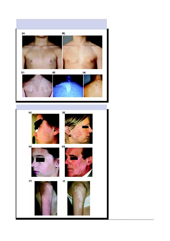
Figure 5: Clichés cliniques d'un patient présentant des lésions lentigineuses sur la face ventrale
(a) et la face dorsale (b) du tronc, correspondant au schéma de dépigmentation chez un patient
atteint de vitiligo segmentaire dans la même région anatomique (b, e). Une photo (d) avec
lumière UV supplémentaire.
Figure 6: Photos cliniques de lésions lentigineuses au visage et au bras (a, c, e) correspondant
au vitiligo segmentaire (b, d, f).