And that’s what Dr. Burns and the AOIAOI researchers are doing with the eye. “We use light coming back out of the eye to tell us all about changes in the lens, the cornea, and the tear film. We measure those things and how they change over time, and we use those programmable mirrors to undo those bendings,” says Dr. Burns. “Adaptive optics allows us to bypass the imperfections of the normal human eye to get about as good an image as it’s possible to get.” People have been able to see the retina since the middle of the nineteenth century, but it’s been “a long slow process of making visibility better and better,” Dr. Burns says. “Around 1990, a lot of forces came together—improved computers, improved electronics for detecting light, improved lasers and light sources for generating light—and this combination of improvements created a revolution in the retinal imaging field.”
As a Ph.D. student in biophysics at The Ohio State University, Dr. Burns studied in an interdisciplinary vision science program in conjunction with Ohio State’s College of Optometry. His postdoctoral study at the University of Chicago was in a unique program through the ophthalmology department that trained basic scientists about eye disease. Through his employment at the Eye and Ear Hospital in Pittsburg, he became fascinated by how diseases like age-related macular degeneration affect the photoreceptors in the retina. But to understand these changes, he needed to clearly see them. Dr. Burns worked at the Eye Research Institute in Boston (later renamed the Schepens Eye Research Institute) for two decades before coming to IU in 2005. He has been studying diseases that affect the retina and refining the imaging technology for viewing them ever since. His retinal imaging research began with a focus on the retinal photoreceptors—the first step in vision—which start the process of turning the light that falls onto the retina into a neural signal that we can then see and interpret.
THE BORDER OF RESEARCH AND PRACTICE
Dr. Burns is regarded as an international leader in the field of retinal function and biomedical imaging, and has “always lived on the border between developing new techniques and using them on patients,” he says. For outstanding contributions to the science of color vision and color imaging systems, he received the Optical Society of America’s Edgar D. Tillyer Award in 2010.
CANARY IN A COAL MINE
“The photoreceptors are very specialized cells,” Dr. Burns says. “They are also some of the most metabolically active cells in the body, which means they’re quite sensitive to being upset
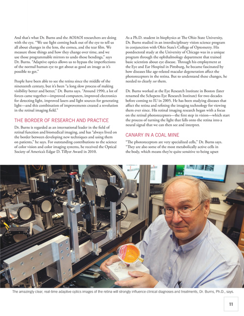
The amazingly clear, real-time adaptive optics images of the retina will strongly influence clinical diagnoses and treatments, Dr. Burns, Ph.D., says.
11